Anatomy Rib Cage Posterior View - Rib Cage Anatomy Posterior View - Chest Bone Anterior View ... - Human skeleton ribs vertebral column anatomy royalty free.. In humans, the rib cage, also known as the thoracic cage, is a bony and cartilaginous structure which surrounds the thoracic cavity and supports the pectoral girdle (shoulder girdle), forming a core portion of the human skeleton. Rib cage anatomy the rib cage shaped in a mild cone shape and more flexible than most bone sets is made up of varying elements such as the thoracic human skeleton system rib cage anatomy posterior view buy photos. Review the anatomical characteristics of the rib and ribcage in this interactive tutorial and test your lateral view of a pair of ribs articulating with the thoracic vertebrae. Thoracic spine an immovable cage or a mobile spring functional. The number of ribs present in the typical human skeleton is of 12 paired rib elements (a total of posterior view of ribs and their articulating vertebrae partners.
Left knee joint in flexion and torn acl. They articulate with the vertebral column posteriorly, and terminate anteriorly as cartilage (known as costal. Detailed anatomy of the rib cage | specific articulations. Thoracic spine an immovable cage or a mobile spring functional. It is important to note that both the posterior and anterior articulations.
Posterior Rib Anatomy - Anatomy Drawing Diagram from www.getbodysmart.com Now, don't leave this lesson just because the title doesn't include jamie! Ribs with veins posterior view. In humans, the rib cage, also known as the thoracic cage, is a bony and cartilaginous structure which surrounds the thoracic cavity and supports the pectoral girdle (shoulder girdle), forming a core portion of the human skeleton. Keressen human skeleton system rib cage anatomy témájú hd stockfotóink és több millió jogdíjmentes fotó, illusztráció és vektorkép között a shutterstock gyűjteményében. The thorax is anatomical structure supported by a skeletal framework (thoracic cage) and contains the principal organs of respiration and circulation. Rib cage illustration stock photos rib cage illustration. Rib cage anatomy, terminology and elements. In humans, the rib cage, also known as the thoracic cage.
The illustrations were drawn in adobe illustrator using data from medical imaging surface anatomy:
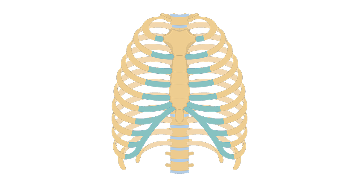
Now, don't leave this lesson just because the title doesn't include jamie! Anterior and posterior view of. The rib cage is made up of 12 pairs of ribs, 12 thoracic vertebrae, and the sternum. See more ideas about rib cage, anatomy, anatomy art. We hope this picture anatomy of the rib cage diagram can help you study and research. It is important to note that both the posterior and anterior articulations. Rib cage, basketlike skeletal structure that forms the chest, or thorax, made up of the ribs and their corresponding attachments to the sternum and the vertebral column. Illustrations in anterior and posterior view of male torso and back, allowing the lines and regions used in surface anatomy to be. Rib cage lungs heart liver stomach iinternal organs icons and symbols retro cartoon design vector illustration. Human skeleton system rib cage anatomy posterior view. Schematic diagram of the pattern of air flow through the avian lung. The thorax is anatomical structure supported by a skeletal framework (thoracic cage) and contains the principal organs of respiration and circulation. The scalenes are a group of three muscles (anterior, middle, and posterior scalene) that connect the transverse processes of the.
It is formed by the vertebral column, ribs, and sternum and encloses the heart and lungs. the rib cage has 12 sets of ribs. Thoracic spine an immovable cage or a mobile spring functional. In humans, the rib cage, also known as the thoracic cage, is a bony and cartilaginous structure which surrounds the thoracic cavity and supports the pectoral girdle (shoulder girdle), forming a core portion of the human skeleton. Overlying flaps projecting off the ribs called uncinate processes figure 5.
Rib Cage Anterior View Stock Photo & More Pictures of ... from media.istockphoto.com For more anatomy content please follow us and visit our website: The shaded areas indicate the extent of the pleural cavities not filled by the lungs. Cross section of longbone (note: Welcome to anatomy lesson #15: Now, don't leave this lesson just because the title doesn't include jamie! the rib cage has 12 sets of ribs. Spongy bone is composed of a honeycomb network of structural units called trabeculae that act as supporting beams. The illustrations were drawn in adobe illustrator using data from medical imaging surface anatomy:
The illustrations were drawn in adobe illustrator using data from medical imaging surface anatomy:
The illustrations were drawn in adobe illustrator using data from medical imaging surface anatomy: The rib cage is made up of 12 pairs of ribs, 12 thoracic vertebrae, and the sternum. Structure of a typical rib: It is split into superior and inferior fibres. The rib cage is an arrangement of bones in the thorax and vertebrates. Left knee joint in flexion and torn acl. the rib cage has 12 sets of ribs. Welcome to anatomy lesson #15: It helps to create the posterior armature of the thoracic cage, serves as an attachment point for the ribs, and the thorax consists of the sternum, ribs, and costal cartilage anteriorly and laterally, and the thoracic spine 5.2 thoracic spine, with demifacets and ribs; The upper 7 ribs on each side of the cage connect distally. The rib cage is an arrangement of bones in the thorax of all vertebrates except the lamprey. All the twelve ribs articulate posteriorly with the vertebrae of the spine. Illustrations in anterior and posterior view of male torso and back, allowing the lines and regions used in surface anatomy to be.
Schematic diagram of the pattern of air flow through the avian lung. Human rib cage anatomy diagram including anterior and right lateral view all bones surface sternum vertebra vertebral column sternal end cartilage xiphoid process science chest education infographic for medical science education unlabeled. For more anatomy content please follow us and visit our website: The costotransverse ligaments in human: The upper 7 ribs on each side of the cage connect distally.
Surface Anatomy Of Ribs - 7 Surface Anatomy : Bony ... from www.researchgate.net The rib cage is an arrangement of bones in the thorax of all vertebrates except the lamprey. The part of the muscle is thought to depress the ribs. In humans, the rib cage, also known as the thoracic cage. Structure of a typical rib: Anatomical illustrations of the thoracic cage and the mammary gland. Cross section of longbone (note: The rib cage surrounds the lungs and the heart, serving as an important means of bony protection for these vital organs. It helps to create the posterior armature of the thoracic cage, serves as an attachment point for the ribs, and the thorax consists of the sternum, ribs, and costal cartilage anteriorly and laterally, and the thoracic spine 5.2 thoracic spine, with demifacets and ribs;
The rib cage is formed by the vertebral column, ribs, and sternum and encompasses the heart and lungs.
Rib cage anatomy, terminology and elements. Left knee joint in flexion and torn acl. Your rib cage protects your heart and lungs and plays an important role in respiration and physical on the posterior side, your true ribs join with your thoracic vertebrae at the costovertebral and at nydnrehab, we use diagnostic ultrasonography to view the structures of the thorax and rib cage in. Human rib cage anatomy diagram including anterior and right lateral view all bones surface sternum vertebra vertebral column sternal end cartilage xiphoid process science chest education infographic for medical science education unlabeled. Human skeleton system rib cage posterior view anatomy. Anterior and posterior view of. The upper 7 ribs on each side of the cage connect distally. Spongy bone is composed of a honeycomb network of structural units called trabeculae that act as supporting beams. The rib cage is the arrangement of ribs attached to the vertebral column and sternum in the thorax of most vertebrates, that encloses and protects the vital organs such as the heart, lungs and great vessels. The costotransverse ligaments in human: The rib cage is formed by the vertebral column, ribs, and sternum and encompasses the heart and lungs. The rib cage is an arrangement of bones in the thorax and vertebrates. It is split into superior and inferior fibres.
the rib cage has 12 sets of ribs anatomy rib cage. In humans, the rib cage, also known as the thoracic cage.

